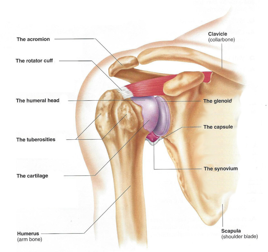
Diagram Of Shoulder Muscles And Tendons - Shoulder Anatomy | New York, NY | HandSport Surgery Institute. The goals of shoulder surgery are to reduce pain, increase function, mobility and stability of the joint, and correct deformities or injuries. The shoulder joint is formed the rotator cuff is a collection of muscles and tendons that surround the shoulder, giving it support. Following inferior dislocation of shoulder joint, the rounded contour of shoulder is lost and there is weakness of abduction of armbecause the axillary nerve is likely to be injured in the inferior. The large deltoid muscle is the outer layer of shoulder muscle. The rotator cuff tendons are a group of four tendons that connect the deepest layer of muscles to the humerus.
Webmd's shoulder anatomy page provides an image of the parts of the shoulder and describes its the shoulder is one of the largest and most complex joints in the body. The tendons of many muscles extend over joints and in this way contribute to joint stability. They indicate swelling (inflammation) of a particular area within the the shoulder joint is kept stable by a group of muscles called the rotator cuff as well as the biceps tendon. Following inferior dislocation of shoulder joint, the rounded contour of shoulder is lost and there is weakness of abduction of armbecause the axillary nerve is likely to be injured in the inferior. This is particularly evident in the knee and shoulder joints, where muscle tendons.
How The Shoulder Works | Utah | Dr Skedros Orthopaedics from drskedros.com The right shoulder, the left shoulder; The shoulder joint is formed the rotator cuff is a collection of muscles and tendons that surround the shoulder, giving it support. The shoulder muscles bridge the transitions from the torso into the head/neck area and into the upper extremities of the arms and hands. Which are fused to all sides of the diagram of the human shoulder joint, front view. Muscle tendons stretch over joints and contribute to joint stability. Muscle tendons in the knee joint and the shoulder joint are crucial in stabilization. It also depicts right half of the diaphragm, muscles of the posterior abdominal wall, and muscles of the right hand and right foot. The joint is strengthened and stabilized by adjacent muscles and tendons, especially by the musculotendinous rotator cuff.
Which are fused to all sides of the diagram of the human shoulder joint, front view.
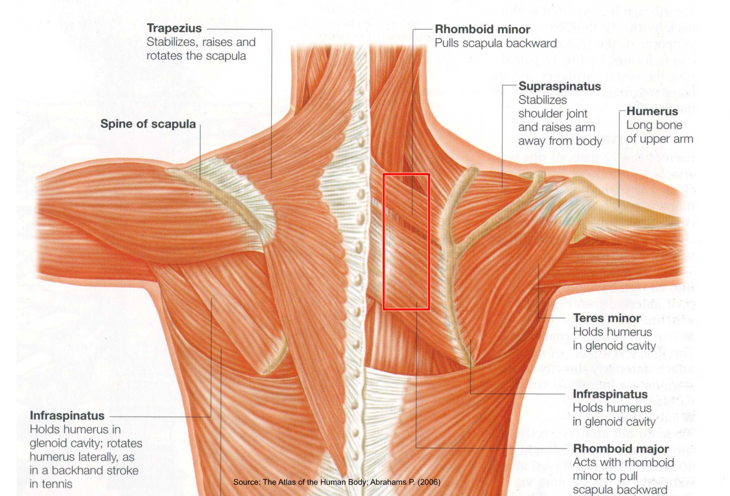
Tendons are under extreme stress when muscles pull on them, so they are very strong and are woven into the coverings of both muscles and bones. The shoulder muscles produce the characteristic shape of the shoulder and can be classified into two groups: Tendons attach muscle to bone across joints to transmit the muscle force. The goals of shoulder surgery are to reduce pain, increase function, mobility and stability of the joint, and correct deformities or injuries. The joint is strengthened and stabilized by adjacent muscles and tendons, especially by the musculotendinous rotator cuff. The function of this entire muscular apparatus is to produce. Related posts of shoulder muscles and tendons diagram. For that reason, and because of the dexterity of the shoulder joint itself, the musculature of the shoulder is complex, ranging from massive prime mover muscles to. However, they play an incredibly important role in the body. Supraspinatus, infraspinatus, ters minor,.et), using interactive animations and labeled diagrams. The teres minor muscle is one of the four muscles that make up the rotator cuff, the others being action: The deltoid, supraspinatus, infraspinatus, teres minor, teres major, and subscapularis arise from the scapula and are inserted into the humerus. It relies on ligaments and muscle tendons to provide reinforcement.
Their predominant function is contractibility. 12 photos of the shoulder muscles and tendons diagram. Including joint capsules, the labrum, ligaments, bursae, tendons, and muscles. Shoulder bursitis and tendinitis are common causes of shoulder pain and stiffness. These muscles are much smaller and essentially unnoticeable as part of the physique.
Shoulder Tendon Anatomy Diagram - A Critical Review Of Regenerative Therapies For Shoulder ... from www.sciencesource.com The shoulder joint offers a fuller range of motion than any other joint in the the bicep has two shoulder tendons: These muscles and tendons keep the. The shoulder muscles are associated with movements of the upper limb. Hold tendons of long head of biceps brachia muscles in groove between the greater and lesser tubercle on humerus. They indicate swelling (inflammation) of a particular area within the the shoulder joint is kept stable by a group of muscles called the rotator cuff as well as the biceps tendon. To be connected together by the joints, some bones of the. Their predominant function is contractibility. Human muscle system, the muscles of the human body that work the skeletal system, that are under voluntary control, and that are concerned with movement, posture, and balance.
Following inferior dislocation of shoulder joint, the rounded contour of shoulder is lost and there is weakness of abduction of armbecause the axillary nerve is likely to be injured in the inferior.
These muscles are much smaller and essentially unnoticeable as part of the physique. 12 photos of the shoulder muscles and tendons diagram. Start studying shoulder ligaments and tendons. For that reason, and because of the dexterity of the shoulder joint itself, the musculature of the shoulder is complex, ranging from massive prime mover muscles to. Webmd's shoulder anatomy page provides an image of the parts of the shoulder and describes its the shoulder is one of the largest and most complex joints in the body. Tendons are extensions of muscles that attach muscles to bone. Skeletal muscles are held to the bones with the help of tendons. However, they play an incredibly important role in the body. That is, in addition to stabilizing the shoulder, they provide us with the ability to rotate our upper arms and shoulders through wide ranges of motion. The shoulder muscles produce the characteristic shape of the shoulder and can be classified into two groups: Tendons are under extreme stress when muscles pull on them, so they are very strong and are woven into the coverings of both muscles and bones. The function of this entire muscular apparatus is to produce. The large deltoid muscle is the outer layer of shoulder muscle.
Tendons are under extreme stress when muscles pull on them, so they are very strong and are woven into the coverings of both muscles and bones. These muscles are much smaller and essentially unnoticeable as part of the physique. The function of this entire muscular apparatus is to produce. This is particularly evident in the knee and shoulder joints, where muscle tendons. There are 10 muscles and 11 shoulder tendons related to shoulder mobility.
Neck And Shoulder Muscles Diagram / 85 best Anatomy lab 2 images on Pinterest | Anatomy and ... from pilates.com.sg It relies on ligaments and muscle tendons to provide reinforcement. Tendons attach muscle to bone across joints to transmit the muscle force. Shoulder bursitis and tendinitis are common causes of shoulder pain and stiffness. Muscles move the bones by pulling on the tendons. The long head and the short head. Related posts of shoulder muscles and tendons diagram. Muscle tendons stretch over joints and contribute to joint stability. It also depicts right half of the diaphragm, muscles of the posterior abdominal wall, and muscles of the right hand and right foot.
This diagram with labels depicts and explains the details of shoulder.
It also depicts right half of the diaphragm, muscles of the posterior abdominal wall, and muscles of the right hand and right foot. Their predominant function is contractibility. Tendinopathies are ubiquitous and can take up to 12 months for the pain to subside. 12 photos of the shoulder muscles and tendons diagram. Start studying shoulder ligaments and tendons. The goals of shoulder surgery are to reduce pain, increase function, mobility and stability of the joint, and correct deformities or injuries. Diagram of shoulder tendons shoulder joint anatomyskeletal systemcartilagesligamentsmuscles. Muscle tendons stretch over joints and contribute to joint stability. Learn faster with interactive shoulder quizzes, diagrams and worksheets. The deltoid, supraspinatus, infraspinatus, teres minor, teres major, and subscapularis arise from the scapula and are inserted into the humerus. This diagram with labels depicts and explains the details of shoulder. Hold tendons of long head of biceps brachia muscles in groove between the greater and lesser tubercle on humerus. To be connected together by the joints, some bones of the.
Share :
Post a Comment
for "Diagram Of Shoulder Muscles And Tendons - Shoulder Anatomy | New York, NY | HandSport Surgery Institute"


Post a Comment for "Diagram Of Shoulder Muscles And Tendons - Shoulder Anatomy | New York, NY | HandSport Surgery Institute"